The acetabulum is the cavity in the pelvis where the femoral head sits: the socket of the hip. Fracturing it means breaking a weight-bearing articular surface, and that sets it apart from almost any other fracture — because here it is not enough for the bone to unite, the surface has to end up smooth.
This is my subspecialty, and the reason I completed a fellowship dedicated to the pelvis, acetabulum and hip. These are uncommon fractures, technically demanding and with a long learning curve; outcomes depend heavily on the experience of the team treating them.
Two very different populations
On one side, the young patient with high-energy trauma: a road traffic collision, a fall from height. There are usually associated injuries, and assessment begins with the whole polytrauma patient, not with the hip.
On the other, the older adult with osteoporotic bone in whom a low-energy fall drives in the medial wall of the acetabulum. The pattern, the options and the goals are different: here the priority is a solution that allows early weight-bearing.
Imaging is half the operation
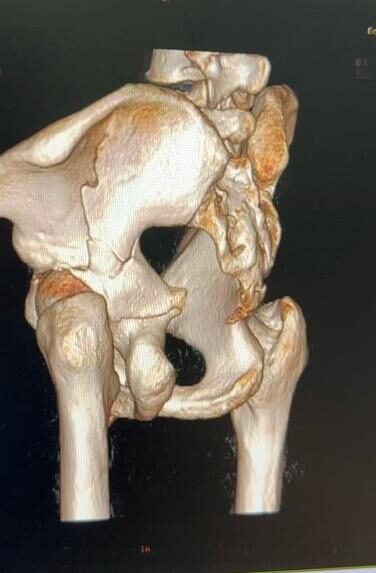
We start with specific radiographs — anteroposterior and the Judet oblique views — but the decisive study is CT with three-dimensional reconstruction. It allows the pattern to be classified according to Judet and Letournel, and that classification is not an academic exercise: it defines which column and which wall are involved, and with that, which approach is required.
Choosing the wrong approach is the error that most determines the outcome, because each route gives access to some structures and not others. Planning happens beforehand, with the images in hand.
Why the reduction has to be millimetric
This is the heart of the injury. A residual step in the articular surface concentrates pressure on the cartilage, and that abnormal pressure eventually produces post-traumatic arthritis years later.
The evidence supports this. A study of long-term patient-reported outcomes after acetabular fracture fixation — with a mean follow-up of 11 years — found that an adequate reduction was associated both with survival of the native hip and with better functional scores. Patients who kept their own hip also reported better function than those who were converted to a total hip replacement.
In plain terms: the quality of the reduction achieved in theatre is what decides whether that hip lasts decades or needs a replacement within a few years.
Treatment options
Fractures that are undisplaced or minimally displaced in non-weight-bearing areas can be treated without surgery, with protected weight-bearing and strict radiographic follow-up to detect any displacement.
Las Displaced fractures are operated on: open reduction and fixation with plates and screws, through whichever approach the pattern demands. The goal is to restore the anatomy of the articular surface.
In older patients with highly comminuted bone, a total hip replacement may be considered from the outset, sometimes combined with posterior column fixation to support the cup. It avoids months of protected weight-bearing and allows faster functional recovery.
Recovery
It usually means several weeks of protected weight-bearing, with early hip mobilisation to avoid stiffness. Follow-up is long, with periodic radiographs, because some complications — femoral head osteonecrosis, post-traumatic arthritis, heterotopic ossification — only appear months or years later.
Dr. Kushner’s approach: in the acetabulum, millimetres matter. Reconstructing the articular surface precisely is what protects the patient’s hip in the long term, and it is one of the areas I dedicated my subspecialty training to.
Cases treated by Dr. Kushner
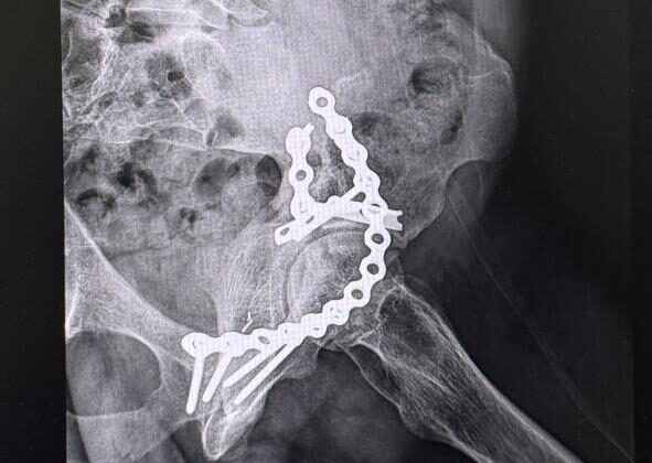
Radiographs from real cases treated by Dr. Kushner (de-identified images, used with consent).
Signs that mean going to the emergency department
- Being unable to bear weight after the injury, or visible deformity of the limb.
- Numbness, loss of strength, or cold or pale toes or fingers below the injury.
- Pain that grows out of proportion and does not respond to analgesia: this may indicate compartment syndrome.
- An open wound over the fractured area.
- Swelling that increases quickly, with tight, shiny skin.
Frequently asked questions
Why does it matter so much that it ends up "perfect"?
Because it is a weight-bearing surface: a step left inside the joint wears the cartilage and brings arthritis forward.
Could I end up needing a replacement?
In some cases, depending on the initial damage. A good reconstruction reduces that risk; when the damage is extensive, a replacement may be the best solution.
Why does this fracture need a specific surgeon?
Because it is the articular surface of the hip: millimetres of step change the long-term prognosis. It requires understanding the patterns, choosing the right approach and reducing precisely.
Will I end up with a replacement?
Not necessarily. A well-achieved anatomical reduction protects the cartilage. In some patterns and in older patients a replacement may be considered from the start, sometimes combined with fixation.
How long without bearing weight?
Usually several weeks of protected weight-bearing, with rehabilitation from early on. The exact period depends on the pattern and on the fixation achieved.
References
- Verbeek DO, et al. Long-term patient reported outcomes following acetabular fracture fixation. Injury. 2018;49(6):1131-1136. DOI
Book your assessment
Book a consultation with Dr. Kushner at COMEDSA. Bring any previous imaging for a complete assessment.
Educational information reviewed by Dr. Michael Kushner Shrem. It does not replace an in-person medical assessment.